

Why Is the Left Ventricle Thicker Than the Right? If you’ve ever looked at a heart diagram, one detail stands out immediately. The left side of the heart looks noticeably bulkier than the right, and that’s not an accident of anatomy.
Why Is the Left Ventricle Thicker Than the Right?
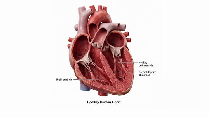
The left ventricle is thicker than the right because it pumps blood through the entire body, a high-resistance system called systemic circulation. The right ventricle only pumps blood to the nearby lungs, a low-pressure path called pulmonary circulation.
More resistance requires more muscular force, and more force requires more muscle mass. That extra muscle is what makes the left ventricular wall so much thicker.
A Quick Refresher on Heart Structure
The heart has four chambers: two atria on top and two ventricles below. Blood enters through the atria and gets pumped out through the ventricles, the heart’s main power source.
The right ventricle sends deoxygenated blood to the lungs through the pulmonary artery. The left ventricle sends oxygenated blood to the rest of the body through the aorta.
Typical Wall Thickness Numbers
| Chamber | Average Wall Thickness | Circulation Type |
|---|---|---|
| Left ventricle | 8–12 mm | Systemic (whole body) |
| Right ventricle | 3–5 mm | Pulmonary (lungs only) |
This gives a rough 3:1 thickness ratio between the two ventricles in a healthy adult heart.
Why Is the Left Ventricle Thicker Than the Right? The Core Reason
1. Distance and Resistance
The left ventricle pushes blood through miles of arteries reaching the brain, limbs, and organs. This systemic circuit carries much higher resistance than the short trip to the lungs.
More resistance means the muscle has to squeeze harder every single beat. Over time, that workload builds thicker, denser muscle tissue.
2. Higher Blood Pressure Requirements
Systolic pressure in the left ventricle typically reaches around 120 mmHg, while the right ventricle usually works at only 25 mmHg. That’s roughly five times more pressure on the left side.
Generating that pressure difference requires a stronger contractile force, which only thicker myocardium can reliably produce.
3. Protecting the Lungs from Damage
The lungs contain extremely delicate capillaries built for gas exchange. If blood arrived there at high pressure, it would damage these thin-walled vessels and cause fluid buildup.
The right ventricle stays thin on purpose, generating just enough force to move blood gently through the lungs without harming them.
4. Muscle Mass and Myocyte Density
Cardiac muscle cells, called myocytes, are more numerous and more densely packed in the left ventricular wall. This isn’t random growth; it’s a direct structural response to workload.
Just like a weightlifter’s muscles grow with resistance training, the heart’s muscle thickens in response to the pressure it repeatedly overcomes.
Left vs. Right Ventricle at a Glance
| Feature | Left Ventricle | Right Ventricle |
|---|---|---|
| Circulation supplied | Systemic (whole body) | Pulmonary (lungs) |
| Average wall thickness | 8–12 mm | 3–5 mm |
| Typical systolic pressure | ~120 mmHg | ~25 mmHg |
| Shape | Thick, cone-like | Thin, crescent-shaped |
| Main outflow vessel | Aorta | Pulmonary artery |
What Happens When the Left Ventricle Gets Too Thick?
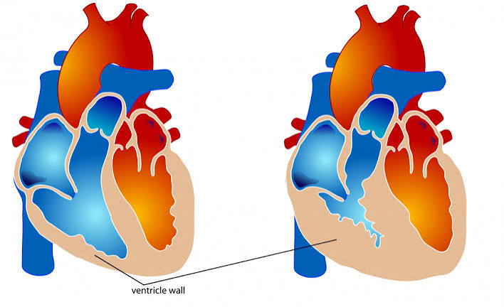
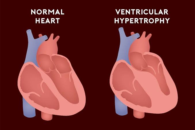
A normally thicker left ventricle is healthy, but excessive thickening signals a problem. This condition is called left ventricular hypertrophy, and it usually develops from chronic high blood pressure or aortic valve disease.
Over time, an overly thick wall can stiffen, reducing the heart’s ability to relax and fill properly between beats. Left untreated, this can progress toward heart failure.
Can the Right Ventricle Also Thicken?

Yes, this is called right ventricular hypertrophy. It typically results from conditions that raise pressure in the lungs, such as chronic lung disease or pulmonary hypertension.
Because the right ventricle is built for low pressure, even a moderate increase in pulmonary resistance can force it to work far harder than normal.
How Doctors Measure Ventricular Wall Thickness
Echocardiogram
An ultrasound of the heart is the most common way to measure wall thickness. It’s non-invasive, quick, and widely available in most hospitals and clinics.
Cardiac MRI
MRI offers highly detailed images and is often used when echocardiogram results are unclear or when very precise measurements are needed.
CT Scan
A cardiac CT scan can also assess wall thickness, particularly alongside imaging for coronary artery disease.
Why This Matters for Heart Health
Understanding ventricular wall thickness helps explain many common heart conditions. Doctors use these measurements to diagnose hypertrophy, monitor heart failure risk, and evaluate valve disease.
For students, this concept also explains a foundational idea in cardiovascular physiology: structure follows function. The heart’s design directly reflects the physical demands placed on each side.
Best Practices for Understanding This Topic
- Always connect wall thickness to workload, not just anatomy
- Remember the ratio is roughly 3:1, not equal on both sides
- Separate normal thickening from pathological hypertrophy
- Use diagrams that show both ventricles side by side for comparison
Mistakes Students Commonly Make
A common mistake is assuming the left ventricle pumps a larger volume of blood than the right. In reality, both ventricles pump the same volume per heartbeat; only the pressure and resistance differ.
Another mistake is confusing wall thickness with chamber size. A thick wall doesn’t always mean a larger chamber; sometimes it means a smaller internal space due to muscle growth.
Frequently Asked Questions (FAQs)
Why is the left ventricle thicker than the right ventricle?
The left ventricle pumps blood through the entire body against high resistance, while the right ventricle only pumps to the lungs at low pressure. That extra workload builds thicker muscle.
Do both ventricles pump the same amount of blood?
Yes. Both ventricles pump nearly identical blood volumes per heartbeat; only the pressure and resistance they work against differ.
What is the normal thickness of the left ventricular wall?
A healthy left ventricular wall typically measures between 8 and 12 millimeters, depending on the individual and measurement method used.
What is left ventricular hypertrophy?
It’s an abnormal thickening of the left ventricular wall, usually caused by long-term high blood pressure or aortic valve disease.
Can the right ventricle become thicker than normal?
Yes, this is called right ventricular hypertrophy, and it’s usually caused by conditions that raise pressure in the lungs.
Is a thicker left ventricle always a bad sign?
No. Some thickness is completely normal and expected. Only excessive or rapid thickening beyond normal ranges is considered a health concern.
How is ventricular wall thickness measured?
Doctors most commonly use echocardiograms, though cardiac MRI and CT scans can also provide detailed measurements.
Why does the right ventricle need to stay thin?
A thin wall keeps pulmonary blood pressure low, protecting the lungs’ delicate capillaries from damage during gas exchange.
What pressure does the left ventricle generate compared to the right?
The left ventricle typically generates around 120 mmHg, roughly five times higher than the right ventricle’s usual 25 mmHg.
Does exercise affect ventricular wall thickness?
Regular cardiovascular exercise can cause healthy, adaptive thickening of the heart muscle, distinct from disease-related hypertrophy.
What shape is the left ventricle compared to the right?
The left ventricle is thick and roughly cone-shaped, while the right ventricle is thinner and more crescent-shaped, wrapping around the left.
Is left ventricular hypertrophy reversible?
In many cases, especially when caused by high blood pressure, effective treatment and lifestyle changes can partially reduce the thickening over time.
Conclusion
At its core, the reason why is the left ventricle thicker than the right comes down to workload. The left side pumps blood across the entire body against high resistance, while the right side handles a short, low-pressure trip to the lungs.
This structural difference isn’t a flaw in design; it’s precise physiological engineering matched to two very different jobs. Understanding it also gives doctors a valuable early clue when something in the cardiovascular system starts to go wrong.
Recognizing normal thickness ranges versus pathological hypertrophy remains one of the most useful diagnostic tools in cardiology today.
